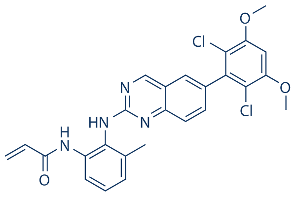
Then compared their prediction performances in detail. To interpret the patterns of morphological AZ 960 features engaged in high-performance models in each differentiation lineage, we selected LASSO regression as a modeling method. Quantitative comparison of morphology and gene expression in prior prediction of differentiation potential. Although morphological information has long been used as an indicator for cellular evaluation, it has remained unclear how descriptive such information really is. To quantitatively compare the performance of morphological and biological information, we directly compared the performances of predictive models using morphological features, gene expression, or both in predicting differentiation potentials from the undifferentiated state. This comparison provides a performance benchmark for our proposed morphology-based cellular potential prediction methodology, enabling complete, non-invasive, daily cellular evaluations that could support or complement evaluations that rely on conventional biomarkers. In our previous study, we found that time courses of morphological features of cultured Nutlin-3 548472-68-0 hBMSCs were informative in the construction of computational models aimed at forecasting future osteogenic differentiation. To evaluate the multiple potentials of hBMSCs in practice, our concept of prediction had to be expanded from single-lineage to multi-lineage differentiation while retaining the ability to adapt to wider cellular variations. However, we hypothesized that in order to predict multi-lineage differentiation potential, informative morphological features and combinations thereof should be optimized for each type of differentiation. In addition, in order to increase the clinical applicability of this approach, our conceptual prediction models had to balance the accuracy with the effort required for model preparation. Therefore, we have set our goal to define the effective construction scheme yielding the optimized prediction performance for each four different types of hBMSC potentials: potential I, osteogenic differentiation rate after 2 weeks of differentiation; potential II, adipogenic differentiation rate after 3 weeks of differentiation; potential III, chondrogenic differentiation rate after 4 weeks of differentiation; and potential IV, PDT of cells after the passages. Our objective prediction  model with the newly developed techniques in this work is illustrated in Fig. S1. To achieve the best prediction models for these objectives, we examined nine patterns of input data usages, which critically change the users’ efforts for data preparation. Model 1 was designed to be the negative control, and Models 2 and 3 were designed to compare gene expression–based predictions compared to morphology-based models. Models 4–9, consisting of five model patterns, were designed to compare morphological feature conversion methods by investigating the various conversion concepts and time-course data usage in morphological features. The comparison of morphological features was deepened by balancing accuracy vs. feasibility of usage in the clinic. Therefore, M-patterns were numbered in the order of higher cost performance, considering the time, cost, and computational memory size involved in the model-construction process.