AbMole Mini-Lecture | BAY 11-7082: A Classic NF-κB Inhibitor in Cancer, Immunology, and Metabolism Research

BAY 11-7082 (BAY 11-7821, M2040, AbMole)  is a widely used NF-κB inhibitor applied in research on inflammatory responses, cancer, and immune regulation. BAY 11-7082 (CAS No.: 19542-67-7) selectively inhibits IκBα phosphorylation in the NF-κB pathway, thereby blocking IκBα degradation and NF-κB nuclear translocation, ultimately suppressing the transcription of NF-κB-dependent genes[1]. In addition to this classical pathway, BAY 11-7082 also inhibits the ubiquitin-specific proteases USP7 and USP21 (IC50: 0.19 μM and 0.96 μM, respectively) and interferes with ubiquitin-conjugating enzyme E2 activity. In RAS-mutant tumor cells, BAY 11-7082 significantly suppresses the growth of NRAS-, KRAS-, and HRAS-mutant tumor cells, an effect validated both in vitro and in mouse xenograft models[1]. Its molecular mechanisms include inhibition of the PI3K-AKT signaling pathway, activation of apoptotic pathways, and downregulation of multiple pro-survival genes [1]. In liver fibrosis research, BAY 11-7082 is used to inhibit activated hepatic stellate cells [2]. BAY 11-7082 also exhibits multiple protective effects in animal models of neurological disorders. For example, it inhibits TNF-α-induced astrocyte dedifferentiation via the NF-κB-Nanog-CD44/Musashi-1 signaling axis[3]; alleviates neuropathy and improves mitochondrial function in diabetic mouse models[4]; and in a postnatal rat model, it inhibits sevoflurane-induced hippocampal pyroptosis and neuroinflammation, preserving synaptic integrity and improving neurocognitive function[5]. In bone metabolism research, BAY 11-7082 is used to validate the regulatory role of the NF-κB pathway in osteogenic differentiation[6]. Thus, BAY 11-7082 is not only an essential tool compound for studying the NF-κB pathway but also demonstrates strong application potential in animal models of cancer, neurodegenerative diseases, fibrotic diseases, and metabolic diseases.

AbMole provides global researchers with high-purity, high-bioactivity inhibitors, cytokines, human monoclonal antibodies, natural products, fluorescent dyes, peptides, compound libraries, antibiotics, and other research reagents, widely cited in numerous publications and patents worldwide.

Case Study

J Inflamm Res. 2021 Mar 17;14:917-928.

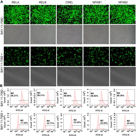
Researchers from the State Key Laboratory of Bioelectronics at Southeast University developed a high-throughput visual screening platform for NF-κB inhibitors using three gene-edited tumor cell lines. Using TALEN and CRISPR technology, they edited five NF-κB family genes (RELA, RELB, CREL, NF-κB1, NF-κB2) in three cell lines (293T, HepG2, and PANC1) to enable ZsGreen fusion expression. BAY 11-7082 (M2040, BAY 11-7821) provided by AbMole was used as an NF-κB inhibitor to validate cellular responsiveness to NF-κB inhibition. In 2014, two inhibitors from AbMole were used in in vivo studies by the Spanish National Center for Cardiovascular Research (CNIC) and Columbia University, leading to publications in Nature and Nature Medicine.

BAY 11-7082 treatment applied to flow cytometry-sorted positive cells [7].

References and Acknowledgments

[1] P. Guruvaiah, R. Gupta, IkappaBalpha kinase inhibitor BAY 11-7082 promotes anti-tumor effect in RAS-driven cancers, Journal of translational medicine 22(1) (2024) 642.

[2] Z. Cheng, F. Li, Y. Qie, et al., Hepatic Stellate Cell Membrane-Camouflaged Nanoparticles for Targeted Delivery of an Antifibrotic Agent to Hepatic Stellate Cells with Enhanced Antifibrosis Efficacy, Nano letters 24(49) (2024) 15827-15836.

[3] Z. Ding, C. Dai, W. Shan, et al., TNF-alpha up-regulates Nanog by activating NF-kappaB pathway to induce primary rat spinal cord astrocytes dedifferentiation, Life sciences 287 (2021) 120126.

[4] L. Sharan, A. Pal, S. S. Babu, et al., Bay 11-7082 mitigates oxidative stress and mitochondrial dysfunction via NLRP3 inhibition in experimental diabetic neuropathy, Life sciences 359 (2024) 123203.

[5] J. Dai, X. Li, C. Wang, et al., Repeated neonatal sevoflurane induced neurocognitive impairment through NF-kappaB-mediated pyroptosis, Journal of neuroinflammation 18(1) (2021) 180.

[6] S. Du, D. Yang, Q. Liu, et al., Ginkgolide B Alleviates LPS-Induced Inhibition of Osteogenic Differentiation in Human Periodontal Ligament Stem Cells by Suppressing the p-IkappaBalpha/NF-kappaB Pathway, Drug design, development and therapy 19 (2025) 8309-8326.

[7] S. Zhang, T. Luo, J. Wang, Stable Cells with NF-κB-ZsGreen Fused Genes Created by TALEN Editing and Homology Directed Repair for Screening Anti-inflammation Drugs, Journal of inflammation research 14 (2021) 917-928.

AbMole Mini-Lecture | Rosiglitazone (BRL 49653): A PPARγ Pathway Agonist and Its Research Applications

Rosiglitazone (BRL 49653, AbMole, M1894) is a thiazolidinedione compound and a high-affinity agonist of peroxisome proliferator-activated receptor gamma (PPARγ). PPARγ is a key, ligand-activated transcription factor within the nuclear receptor superfamily, regulating gene expression involved in energy metabolism and cell differentiation. Rosiglitazone binds directly to the PPARγ ligand-binding domain to exert its biological effects by modulating downstream target genes [1, 2].


At the molecular level, Rosiglitazone (CAS No.: 122320-73-4) regulates inflammatory signaling like NF-κB and metabolic pathways including glucose transport and lipogenesis. For example, it reduces p65 phosphorylation and upregulates IκBα expression—an anti-inflammatory effect abolished upon PPARγ knockout, confirming its dependence on PPARγ activation. Recent studies also identify Rosiglitazone as a ligand for retinoid X receptor α (RXRα), regulating gene transcription via an RXRα-dependent pathway [3].


In cellular assays, Rosiglitazone demonstrates pleiotropic effects:
1.Inhibits proliferation: Suppresses cell cycle and induces apoptosis in a dose- and time-dependent manner, significantly reducing migration in 5637 and T24 cells [4].
2.Regulates mitochondrial function: Protects mitochondria, promotes oxidative phosphorylation, and increases intracellular ATP levels—effects reversed by the PPARγ antagonist GW9662 (AbMole, M2748) [5].
3.Modulates macrophage polarization: Inhibits M1 while promoting M2 macrophage polarization and enhances microglial phagocytosis, mediated by the PPARγ/CD36 axis [6].
4.Induces differentiation: Promotes lipid accumulation and differentiation of 3T3-L1 preadipocytes into adipocytes [7].


In vivo, Rosiglitazone reduces neurodamage in a mouse intracerebral hemorrhage model by inhibiting apoptosis via the PPARγ/JNK/STAT3 axis[8]. It also alleviates fibrosis progression in a mouse pulmonary fibrosis model by suppressing p38 MAPK phosphorylation [9].


AbMole provides global researchers with high-purity, high-bioactivity inhibitors, cytokines, human monoclonal antibodies, natural products, fluorescent dyes, peptides, compound libraries, antibiotics, and other research reagents, widely cited in numerous publications and patents worldwide.

Case Study
Adv Sci (Weinh). 2023 May;10(15):e2207224.
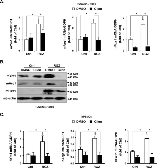
In the above study, researchers at Chongqing Medical University investigated how hypoxia drives biomaterial-induced heterotopic ossification (HO) by modulating macrophage polarization and osteoclastogenesis. The key finding is that the hypoxic environment promotes M2 polarization and lipid accumulation in macrophages via activation of hypoxia-inducible factor-1α (HIF-1α), which subsequently leads to macrophage fusion and osteoclast formation. The osteoclasts then secrete factors (such as CTHRC1 and S1P) that induce osteogenic differentiation of mesenchymal stem cells, ultimately resulting in heterotopic ossification. This mechanism provides new insights for the design of bone repair materials. Rosiglitazone (BRL 49653, AbMole, M1894), an M2 macrophage activator, was used as a positive control in this study to confirm the role of macrophage polarization in the above model.


In 2014, two AbMole inhibitors were used in in vivo studies by the Spanish National Center for Cardiovascular Research (CNIC) and Columbia University, leading to publications in Nature and Nature Medicine.


Cilengitide partially inhibited rosiglitazone-induced M2 polarization.


*The products mentioned herein are for research use only.

References and Acknowledgements
[1] C. Hu, H. L. Keen, K. T. Lu, et al., Retinol-binding protein 7 is an endothelium-specific PPARgamma cofactor mediating an antioxidant response through adiponectin, JCI insight 2(6) (2017) e91738.
[2] Q. Mu, Q. He, H. Zhou, et al., Rosiglitazone Promotes Microglial Distribution via Activation of PPARgamma and CD36 in the ICH Rat Model, Neuro endocrinology letters 45(2) (2024) 96-106.
[3] F. Huang, Y. Li, J. Chen, et al., Rosiglitazone binds to RXRalpha to induce RXRalpha tetramerization and NB4 cell differentiation, Biochemical and biophysical research communications 530(1) (2020) 160-166.
[4] X. Xu, J. Wang, H. Jiang, et al., Rosiglitazone induces apoptosis on human bladder cancer 5637 and T24 cell lines, International journal of clinical and experimental pathology 10(10) (2017) 10197-10204.
[5] J. M. Ortiz-Rodriguez, C. Balao da Silva, J. Masot, et al., Rosiglitazone in the thawing medium improves mitochondrial function in stallion spermatozoa through regulating Akt phosphorylation and reduction of caspase 3, PloS one 14(7) (2019) e0211994.
[6] Q. Mu, L. Wang, H. Hang, et al., Rosiglitazone pretreatment influences thrombin-induced phagocytosis by rat microglia via activating PPARgamma and CD36, Neuroscience letters 651 (2017) 159-164.
[7] Y. Wang, Z. Yang, Y. Li, et al., Impact of Rosiglitazone on Subdermal Adipose Tissue Growth and Lipid Droplet Formation: An In Vitro and In Vivo Study, Aesthetic plastic surgery (2025).
[8] C. Chao, Y. Li, Q. Li, et al., Inhibitory effect and mechanism of Rosiglitazone on M1 type polarization of central microglia in intracerebral hemorrhage mice based on JNK/STAT3 signaling pathway, Brain and behavior 13(12) (2023) e3275.
[9] H. Zhang, L. You, M. Zhao, Rosiglitazone attenuates paraquat-induced lung fibrosis in rats in a PPAR gamma-dependent manner, European journal of pharmacology 851 (2019) 133-143.

AbMole Mini-Lecture | Vorinostat (SAHA, MK0683): Applications of a Histone Deacetylase (HDAC) Inhibitor in Epigenetics, Cancer, and Neurobiology

Vorinostat (SAHA, MK0683, AbMole, M1780) is a broad-spectrum, pan-histone deacetylase inhibitor (HDACi). Belonging to the succinylhydroxamic acid class of compounds, it directly binds to and inhibits HDAC activity, leading to the accumulation of acetylated histones, thereby regulating gene expression and affecting multiple signaling pathways. Additionally, Vorinostat (MK0683) exhibits bioactivities including cell cycle arrest, apoptosis, and inhibition of angiogenesis. Studies have shown that Vorinostat bidirectionally regulates stress response genes (e.g., sod-3, hsp-16.2, skn-1). It upregulates these genes at low concentrations but exhibits inhibitory or neutral effects at high concentrations (10 μM), demonstrating dose-dependent epigenetic regulatory properties [1]. Vorinostat also induces cell cycle arrest and programmed cell death by downregulating the Akt signaling pathway [2]. Furthermore, Vorinostat downregulates multiple epigenetic regulatory enzymes (e.g., EZH2, SUV39H1/2, DOT1L), indicating its broad impact on the epigenetic regulatory network [3].

In research applications, Vorinostat (SAHA) has shown multifaceted potential. First, it inhibits the proliferation of various tumor cells, including colon cancer cells (HCT116, HT29) [4], pancreatic cancer cells (AsPC-1) [5], hepatocellular carcinoma cells (LCL-PI 11) [5], and head and neck squamous cell carcinoma (HNSCC) cells [6]. Vorinostat is also used to inhibit parasites, with the mechanism similarly involving HDAC inhibition in protozoan cells [7]. Notably, the activity status of tissue transglutaminase 2 (TG2) affects cellular sensitivity to Vorinostat. Inhibition of TG2 activity enhances the anti-proliferative effect of Vorinostat, whereas TG2 overexpression confers resistance, identifying TG2 as a key target in Vorinostat tolerance mechanisms [8]. In the field of neuroprotection, Vorinostat reverses amyloid β protein -induced neural damage, an activity involving regulation of the AKT-MDM2-p53 pathway [9].

AbMole provides global researchers with high-purity, high-bioactivity inhibitors, cytokines, human monoclonal antibodies, natural products, fluorescent dyes, peptides, compound libraries, antibiotics, and other research reagents, widely cited in numerous publications and patents worldwide.

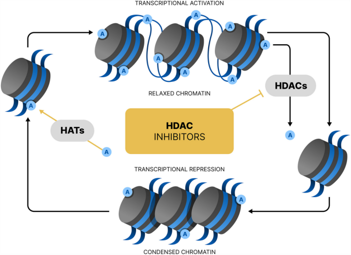
Transcriptional activity of histone deacetylase (HDAC) inhibitors[10].

Case Study

BMC Cancer. 2016 Nov 7;16(1):857.

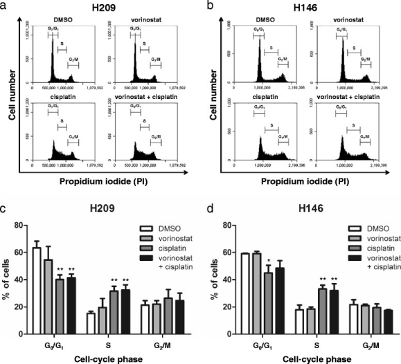
In this study, researchers investigated the inhibitory effect of Vorinostat on small cell lung cancer (SCLC) and the development of combination therapy strategies. Through in vitro (H209, H146 cell lines) and in vivo (H209 xenograft nude mouse model) experiments, the study demonstrated that Vorinostat (an HDAC inhibitor), when combined with Cisplatin or with Etoposide (VP-16-213), more significantly inhibited tumor cell viability, induced apoptosis (via caspase-3 activation and PARP cleavage), and induced S-phase cell cycle arrest compared to single-agent treatments. The combination also increased acetylation levels of histone H3 and α-tubulin while persistently inhibiting thymidylate synthase (TS) expression. The core research compound, Vorinostat (SAHA, MK0683, AbMole, M1780), was provided by AbMole. In 2014, two inhibitors from AbMole were used in in vivo studies by the Spanish National Center for Cardiovascular Research (CNIC) and Columbia University, leading to research publications in Nature and Nature Medicine.

Cell cycle progression analysis of vorinostat in combination with cisplatin in SCLC cells[11].

References and Acknowledgements

[1] S. Huang, H. Shi, Z. Shi, et al., Vorinostat, a potential hormetin, extends lifespan and enhances stress resistance via the SKN-1 pathway in Caenorhabditis elegans, Biogerontology 26(3) (2025) 97.
[2] S. Takeuchi, T. Hase, S. Shimizu, et al., Phase I study of vorinostat with gefitinib in BIM deletion polymorphism/epidermal growth factor receptor mutation double-positive lung cancer, Cancer science 111(2) (2020) 561-570.
[3] V. Maksimova, J. Makus, V. Popova, et al., Histone Methyltransferases as a New Target for Epigenetic Action of Vorinostat, Biochemistry. Biokhimiia 88(7) (2023) 968-978.
[4] M. Yousefian, M. Hashemi, V. Eskandarpour, et al., New indolin-2-ones, possessing sunitinib scaffold as HDAC inhibitors and anti-cancer agents with potential VEGFR inhibition activity; design, synthesis and biological evaluation, Bioorganic chemistry 156 (2025) 108231.
[5] M. Sanaei, F. Kavoosi, Effect of vorinostat on INK4 family and HDACs 1, 2, and 3 in pancreatic cancer and hepatocellular carcinoma, Research in pharmaceutical sciences 16(3) (2021) 260-268.
[6] N. Tanaka, A. A. Patel, L. Tang, et al., Replication Stress Leading to Apoptosis within the S-phase Contributes to Synergism between Vorinostat and AZD1775 in HNSCC Harboring High-Risk TP53 Mutation, Clinical cancer research : an official journal of the American Association for Cancer Research 23(21) (2017) 6541-6554.
[7] H. Li, E. M. Galon, S. Ji, et al., In vitro screening of compounds from the Food and Drug Administration-approved library identifies anti-Babesia gibsoni activity of idarubicin hydrochloride and vorinostat, Parasitology international 96 (2023) 102774.
[8] C. Carbone, E. Di Gennaro, G. Piro, et al., Tissue transglutaminase (TG2) is involved in the resistance of cancer cells to the histone deacetylase (HDAC) inhibitor vorinostat, Amino acids 49(3) (2017) 517-528.
[9] J. Meng, Y. Li, M. Zhang, et al., A combination of curcumin, vorinostat and silibinin reverses Aβ-induced nerve cell toxicity via activation of AKT-MDM2-p53 pathway, PeerJ 7 (2019) e6716.
[10] R. Parveen, D. Harihar, B. P. Chatterji, Recent histone deacetylase inhibitors in cancer therapy, Cancer 129(21) (2023) 3372-3380.
[11] C. H. Pan, Y. F. Chang, M. S. Lee, et al., Vorinostat enhances the cisplatin-mediated anticancer effects in small cell lung cancer cells, BMC cancer 16(1) (2016) 857.

MEK-ERK pathway inhibition underlies the cellular effects of 4-hydroxytamoxifen

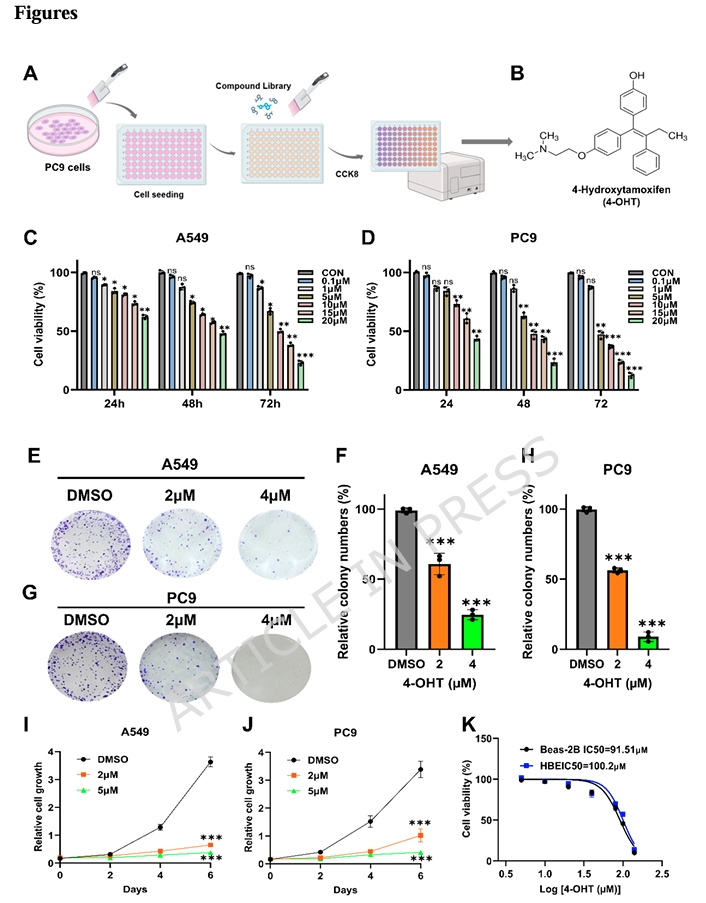
4-Hydroxytamoxifen has spent decades in the shadow of its parent compound, largely catalogued as an estrogen receptor antagonist generated by hepatic metabolism. A recent report in Scientific Reports pulls it into an entirely different spotlight, mapping its effects on MAPK signaling and lipid metabolism in human epithelial cell lines. The investigation began with a high-throughput screen of a human endogenous metabolite library in PC9 cells, where 4-OHT emerged as a potent viability suppressor. Follow-up validation in A549 cells confirmed the initial observation, setting the stage for a mechanistic dissection that links MEK-ERK phosphorylation status to intracellular fatty acid abundance and cellular behavior.

The first layer of evidence came from quantitative proliferation assays. Using a CCK-8 cell counting kit supplied by AbMole, the team measured viability across a concentration gradient spanning 0.1 to 20 μM over 24, 48, and 72 hours. The resulting IC₅₀ curves were steep and time-dependent: A549 cells registered 20.64 μM at 24 hours, dropping to 9.76 μM by 72 hours, while PC9 cells proved even more responsive, moving from 19.41 μM to 6.34 μM over the same interval. What distinguished this profile from generic cytotoxicity was the selectivity window. Non-transformed bronchial epithelial lines HBE and Beas-2B registered IC₅₀ values near 100 μM, indicating that 4-OHT discriminates between transformed and non-transformed phenotypes rather than indiscriminately suppressing all dividing cells. Colony formation assays and crystal violet staining reinforced the CCK-8 data, showing that clonogenic potential and cumulative growth both collapse under sustained 4-OHT exposure in a manner that follows the same dose-response logic.

Beyond simply slowing division, 4-OHT altered how these cells interact with their surroundings. Wound healing assays showed dramatically reduced closure rates at 5 μM, and transwell migration counts dropped in a dose-dependent fashion. Western blotting offered a molecular explanation for the motility defect: mesenchymal markers vimentin and N-cadherin fell, while epithelial E-cadherin rose. This reversal of epithelial-mesenchymal transition suggests 4-OHT does not merely block proliferation but actively reshapes cellular architecture toward a less motile, more adherent state. Parallel flow cytometry with Annexin V-FITC and propidium iodide revealed a second, distinct mechanism—programmed cell death. The fraction of apoptotic cells climbed steadily with dose, indicating that growth suppression is coupled to activation of cell-intrinsic death programs rather than being a simple consequence of metabolic starvation.

To understand the transcriptional logic driving these phenotypes, the researchers turned to RNA sequencing. Twenty-four hours of 20 μM 4-OHT in A549 cells yielded 701 differentially expressed genes, with 416 upregulated and 285 downregulated. KEGG pathway enrichment placed the MAPK signaling cascade among the most significantly altered pathways. More specifically, a coordinated cluster of fatty acid metabolism genes—including FASN, FADS2, ACAT2, ACSS2, and HMGCR—showed consistent downregulation. Gene set enrichment analysis independently flagged fatty acid degradation and related metabolic processes as significantly altered. These transcriptional shifts pointed toward a metabolic bottleneck rather than a single-gene off-target effect, suggesting that 4-OHT remodels the lipid biosynthetic program at the level of gene expression.

Untargeted metabolomics confirmed the prediction with remarkable clarity. Principal component analysis cleanly separated control and 4-OHT-exposed populations, and orthogonal partial least squares-discriminant analysis reinforced the distinction. Lipid and lipid-like molecules constituted the largest category of depleted metabolites, with numerous fatty acid species showing marked reduction. Direct colorimetric quantification of intracellular free fatty acids showed dose-responsive drops in both A549 and PC9 cells, paralleling the transcriptomic downregulation of synthetic enzymes. The metabolic and transcriptional data converged on a straightforward model: 4-OHT starves cells of lipid building blocks by suppressing de novo fatty acid synthesis at multiple enzymatic steps.

The MAPK connection emerged cleanly from Western blotting. Phosphorylated MEK and phosphorylated ERK both declined sharply after 4-OHT exposure, while total protein levels remained unchanged. This pattern implicates post-translational regulation—specifically, inhibition of kinase activation rather than reduced expression or stability. Since MAPK signaling is known to feed into lipid biosynthetic programs through transcriptional control of rate-limiting enzymes like FASN, the researchers designed rescue experiments to test whether forced reactivation of the pathway could override the metabolic phenotype. Combined exposure with C16-PAF, a selective MAPK activator, partially restored p-MEK and p-ERK levels in both A549 and PC9 cells. More importantly, it rescued free fatty acid content, effectively reversing the lipid depletion caused by 4-OHT alone. Functionally, C16-PAF also restored proliferative capacity in crystal violet assays and migratory behavior in transwell and wound healing experiments. The rescue was partial rather than complete, which is expected given that 4-OHT likely engages multiple parallel mechanisms, but the directionality was unambiguous.

To further tighten the causal inference, the team combined 4-OHT with U0126, a selective MEK1/2 inhibitor. This combination produced deeper suppression of MEK-ERK phosphorylation than either molecule alone, and further accentuated the downregulation of fatty acid metabolism genes. The epistasis is clear: 4-OHT sits upstream of MEK-ERK activation, and the lipid metabolic phenotype is a downstream consequence of reduced pathway flux. The fact that a MEK inhibitor synergizes with 4-OHT rather than duplicating its effect at a ceiling suggests that 4-OHT may impinge on the MAPK axis at a level distinct from MEK itself, or that it simultaneously modulates parallel inputs that converge on ERK.

In vivo validation used a syngeneic C57BL/6J mouse model receiving subcutaneous implantation of LLC cells. Once palpable subcutaneous masses reached approximately 100 mm³, mice received daily intraperitoneal administration of 4-OHT at 40 mg/kg for seven consecutive days. The compound substantially reduced subcutaneous mass volume and weight without triggering body weight loss or hepatorenal histopathology in H&E-stained sections. Immunohistochemical Ki-67 staining fell markedly, while TUNEL positivity rose, mirroring the in vitro proliferation suppression and apoptosis induction. These observations confirm that the MAPK-lipid mechanism operates in an intact physiological setting and is not an artifact of two-dimensional culture.

Several aspects of this study merit emphasis for researchers working at the intersection of signal transduction and metabolism. First, the selectivity for transformed over non-transformed epithelial cells indicates a discrimination window that is not obvious from standard cytotoxicity profiles. Second, the rescue experiments with C16-PAF and U0126 go beyond simple correlation, functionally demonstrating that MEK-ERK phosphorylation status is rate-limiting for both the metabolic and behavioral phenotypes. Third, the integration of transcriptomics and untargeted metabolomics provides a multi-omic anchor for the MAPK-lipid axis that might otherwise be dismissed as a secondary or compensatory effect.

There are limits, of course. The study focused on two related epithelial lines, and the precise transcription factor intermediaries linking MAPK suppression to reduced FASN and HMGCR expression remain uncharacterized. Whether the same mechanism operates in mesenchymal or hematopoietic contexts is an open question. Additionally, the pharmacokinetic behavior of 4-OHT in murine systems was not profiled, so the relationship between the in vitro IC₅₀ and the in vivo dose remains correlative rather than quantitatively linked. Nevertheless, the work establishes 4-OHT as a probe for dissecting the intersection of MAPK signaling and lipid homeostasis, and it positions MEK-ERK-mediated fatty acid synthesis as a central node through which 4-OHT coordinates proliferation, motility, and survival decisions in epithelial cell models.

AbMole Product Integration in This Study

Product: CCK-8 Cell Counting Kit (AbMole, USA)

Application: Quantitative assessment of cell viability and proliferation across human epithelial cell lines to establish dose-response relationships and IC₅₀ values for 4-hydroxytamoxifen.

Experimental Details:

  • Cell seeding density: 3,000 cells per well in 96-well plates
  • Compound exposure: 4-OHT at concentrations ranging from 0.1 to 20 μM for 24, 48, and 72 hours
  • Assay execution: CCK-8 reagent added per manufacturer protocol; absorbance read at 450 nm using a microplate reader
  • Analysis: IC₅₀ values calculated via GraphPad Prism 10.0 software
  • Cell lines profiled: A549, PC9, HBE, and Beas-2B

Key Findings Enabled by AbMole CCK-8:

  • A549 IC₅₀ at 72 h: 9.76 ± 1.9 μM; PC9 IC₅₀ at 72 h: 6.34 ± 2.7 μM
  • Non-transformed HBE and Beas-2B bronchial epithelial cells exhibited IC₅₀ values near 100 μM, revealing a roughly tenfold selectivity window
  • Dose- and time-dependent viability reduction confirmed across both epithelial lines, with tamoxifen showing comparable IC₅₀ profiles

Meta Description: Mechanistic analysis of 4-OHT-mediated suppression of MEK-ERK phosphorylation and fatty acid metabolism reprogramming, quantified with AbMole CCK-8 assays in human epithelial cell lines.

Target Keywords: 4-hydroxytamoxifen, MAPK signaling, MEK, ERK, lipid metabolism, fatty acid synthesis, FASN, EMT, AbMole, CCK-8, cell viability

The Sirt3-ERS-Mitophagy Axis: How 3-TYP Intervenes in Cardiomyocyte Stress Responses

Sirt3 has long been positioned as a guardian of mitochondrial integrity, a deacetylase that fine-tunes oxidative phosphorylation and limits reactive oxygen species emission under physiological stress. Located primarily in the mitochondrial matrix, it targets dozens of lysine residues on metabolic enzymes, ostensibly preserving ATP output and organelle fidelity. Yet the assumption that more Sirt3 always equals better cellular health has begun to fray. A recent report in Journal of Molecular Histology adds weight to the counter-narrative, showing that Sirt3 expression surges under combined hypertensive, hyperglycemic, and lipid-excess conditions in rat cardiomyocytes, and that pharmacological suppression of this enzyme—achieved with the selective inhibitor 3-TYP supplied by AbMole—restores architectural and molecular homeostasis. The study pivots attention away from simple sirtuin elevation strategies and toward a more nuanced understanding of the Sirt3-endoplasmic reticulum stress-mitophagy axis as a dynamically regulated network.

The investigators used spontaneously hypertensive rats shifted to a high-fat diet at twenty-four weeks, followed by a single intraperitoneal streptozotocin injection to introduce glucose dysregulation. The result was a multi-hit metabolic challenge: fasting glucose climbed above 16.7 mM, lipids rose, blood pressure remained elevated, and exercise tolerance declined. Control groups included normotensive Wistar-Kyoto rats and untreated spontaneously hypertensive rats, creating a gradient of metabolic insult severity.

In spontaneously hypertensive rats with only the hypertensive background, Sirt3 protein levels in left ventricular tissue actually decreased relative to normotensive controls. This aligns with older literature suggesting hemodynamic stress alone suppresses mitochondrial sirtuin expression. However, when high-fat feeding and streptozotocin were layered on top, Sirt3 expression flipped—rising significantly above baseline. This paradoxical upregulation coincided with structural remodeling: increased heart weight-to-body weight ratios, enlarged cardiomyocyte cross-sectional areas by wheat germ agglutinin staining, disordered sarcomere alignment, and collagen accumulation by Masson and Sirius red microscopy. Pulmonary wet-to-dry weight ratios also increased. The echocardiographic profile showed preserved ejection fraction but elevated left ventricular wall thickness and reduced E/A ratios, pointing toward diastolic stiffening. Serum brain natriuretic peptide, an indicator of myocardial wall stress, climbed markedly.

To determine whether the Sirt3 surge was adaptive or pathogenic, the team administered 3-TYP, a Sirt3-selective inhibitor obtained from AbMole under catalog number M8978, at 50 mg/kg body weight via intraperitoneal injection every other day for nine doses beginning at twenty-seven weeks. The compound was prepared according to supplier specifications, with dosing converted from human equivalents based on body surface area ratios. The outcomes were unambiguous. Rats receiving 3-TYP showed reduced brain natriuretic peptide, improved E/A ratios, and attenuated wall thickening. Histologically, cardiomyocyte cross-sectional area decreased, collagen deposition diminished, and pulmonary congestion indices normalized. The inhibitor restructured the molecular environment of the myocardium in a way that suggested Sirt3 elevation was actively sustaining the stress response rather than resolving it.

The link between Sirt3 and endoplasmic reticulum stress has been proposed previously, but this study delivers rigorous confirmation. Under metabolic stress, the ER faces increased protein folding load. When capacity is exceeded, the unfolded protein response activates through sensors including PERK. Here, GRP78 and PERK both rose sharply in metabolically challenged rats and fell after 3-TYP administration. This indicates that Sirt3 elevation in this context sustains PERK phosphorylation and downstream programs rather than alleviating proteostatic burden.

Ultrastructural analysis via transmission electron microscopy provided compelling evidence. In metabolically challenged rats, cardiomyocytes displayed severe organelle pathology: mitochondria were massively swollen with discontinuous cristae, the ER showed dilated tubules, and autophagosomes containing mitochondrial fragments were abundant. Mitochondrial morphology was scored on a five-grade scale, and the distribution skewed heavily toward severe damage. Following 3-TYP administration, average mitochondrial cross-sectional area and Feret diameter decreased, ER lumen dilation receded, and high-damage scores dropped. Western blotting reinforced these images: GRP78, PERK, Parkin, and LC3-II all decreased after 3-TYP, indicating that Sirt3 inhibition simultaneously dampened ER stress and mitophagy flux.

The in vitro experiments translated these observations into a controlled cellular context. AC16 human ventricular cardiomyocytes were challenged with angiotensin II and high glucose to simulate the metabolic milieu. Angiotensin II was sourced from AbMole under catalog number M6240 and applied at 1 μM for 48 hours, either alone or with 30 mM glucose. Ang II alone reduced Sirt3 expression, consistent with hemodynamic stress suppressing the enzyme. However, the combination of Ang II and high glucose increased Sirt3, mirroring the animal model. Crystal violet staining revealed cellular hypertrophy in the dual-challenge group, and atrial natriuretic peptide protein levels rose significantly. When Sirt3 was knocked down using small interfering RNA, the hypertrophic phenotype collapsed. Cell cross-sectional area returned toward control levels, and atrial natriuretic peptide normalized. This genetic confirmation paralleled the chemical inhibition results with 3-TYP, strengthening the argument that Sirt3 elevation in multi-factorial metabolic stress is causally linked to adverse remodeling.

Fluorescence microscopy offered additional mechanistic granularity. ER-Tracker blue staining showed nonuniform fluorescence and cavity cavitation in cells exposed to Ang II plus high glucose, while MitoTracker red revealed depressed mitochondrial membrane potential. Sirt3 silencing restored uniform ER morphology and improved mitochondrial membrane potential. Western blotting confirmed the pattern: GRP78, PERK, C/EBP homologous protein, Parkin, LC3-II, and Beclin1 all increased under combined metabolic stress and decreased after Sirt3 knockdown. The CHOP result merits attention because this transcription factor bridges ER stress sensing to apoptotic execution. Its suppression following Sirt3 inhibition implies the pathway feeds into programmed cell death machinery under sustained metabolic challenge.

Synthesizing these data yields a model centered on the Sirt3-ERS-mitophagy triad. Under isolated hemodynamic stress, Sirt3 declines—possibly as an adaptive energy-sparing response. When hyperglycemia and hyperlipidemia are superimposed, Sirt3 rises sharply and drives excessive ER stress and mitophagy. This overactivation strips cardiomyocytes of essential mitochondrial mass and destabilizes ER proteostasis, generating a feed-forward loop of organelle dysfunction. Mitophagy shifts from quality-control to destructive self-consumption. The observation that Beclin1 and Parkin both rise under stress and fall after Sirt3 suppression supports the interpretation that Sirt3 acts as a rheostat for autophagic flux. By applying 3-TYP, the researchers uncoupled this loop, allowing cardiomyocytes to re-establish mitochondrial membrane potential and normalize stress protein expression.

This work carries broader implications for cellular stress biology. It underscores that sirtuin function is not monotonic. The same enzyme can be protective in one context and maladaptive in another depending on stress composition. Prior studies showing Sirt3-mediated protection used acute oxidative insults, whereas this work operates in chronic nutrient-excess environments. The discrepancy suggests Sirt3’s role pivots on whether the challenge is oxidative, hemodynamic, or metabolic. Second, the study highlights the danger of excessive mitophagy. While basal mitophagy clears damaged organelles, hyperactivation depletes functional networks and triggers compensatory remodeling. Sirt3 inhibition suppresses both Parkin recruitment and LC3-II lipidation, pointing to a regulatory node that determines whether mitochondrial turnover remains homeostatic or becomes catabolic.

Technically, the investigation benefits from converging genetic and pharmacological evidence. The siRNA experiments in AC16 cells recapitulate the 3-TYP results in rats, reducing concerns about off-target effects. The AbMole reagents—3-TYP for in vivo Sirt3 inhibition and Ang II for in vitro metabolic stress induction—provided standardized, batch-consistent perturbations. Dosage parameters were explicitly reported: 50 mg/kg for 3-TYP delivered intraperitoneally every other day, and 1 μM Ang II for 48 hours in serum-supplemented DMEM. Such specificity enhances reproducibility.

Several questions remain. The precise intermediates linking Sirt3 deacetylase activity to PERK phosphorylation have not been fully mapped. Possibilities include direct acetylation of ER membrane proteins, indirect modulation through mitochondrial ROS feedback, or crosstalk at mitochondria-associated ER membranes. Additionally, the study did not explore whether PGC-1α or FOXO1 mediate the observed phenotype. Dose-response refinement of 3-TYP and temporal dynamics of Sirt3 expression also remain open.

In conclusion, this study reframes Sirt3 as a context-dependent modulator of cardiac cellular stress. Under combined hypertensive and metabolic challenge, Sirt3 elevation paradoxically sustains excessive endoplasmic reticulum stress and mitophagy, driving organelle dysfunction and cellular remodeling. The selective Sirt3 inhibitor 3-TYP, alongside the Ang II stressor used in parallel cell culture experiments, enabled precise dissection of this axis and demonstrated that suppression—not augmentation—of Sirt3 activity restores molecular homeostasis. The work serves as a reminder that sirtuin biology resists simple gain-versus-loss narratives; the metabolic milieu dictates whether these enzymes function as cellular buffers or accelerants of stress-induced remodeling.

AbMole Product Integration in This Study

Product 1: 3-TYP (AbMole, catalog M8978)

Application: Selective Sirt3 inhibitor for in vivo interrogation of Sirt3-dependent signaling in a rat model of compounded metabolic stress.

Experimental Details:

  • Dose: 50 mg/kg body weight
  • Route: Intraperitoneal injection
  • Schedule: Every other day for nine total administrations, initiated at 27 weeks of age
  • Preparation: Dosing converted from human equivalent calculations based on body surface area ratios; prepared and handled per supplier instructions
  • Model: Spontaneously hypertensive rats previously subjected to high-fat diet and streptozotocin challenge

Key Findings Enabled by 3-TYP:

  • Marked reduction in serum brain natriuretic peptide and restoration of E/A ratios
  • Attenuation of left ventricular wall thickening and cardiomyocyte cross-sectional area
  • Significant reduction in collagen deposition by Masson and Sirius red staining
  • Normalization of pulmonary congestion indices
  • Downregulation of ER stress markers (GRP78, PERK) and mitophagy proteins (Parkin, LC3-II)
  • Restoration of mitochondrial and ER ultrastructure by transmission electron microscopy

Product 2: Angiotensin II (AbMole, catalog M6240)

Application: In vitro metabolic stressor for AC16 human ventricular cardiomyocytes.

Experimental Details:

  • Concentration: 1 μM
  • Duration: 48 hours
  • Media: DMEM supplemented with 10% fetal bovine serum
  • Conditions: Applied alone or in combination with high glucose (30 mM) to simulate multi-factorial metabolic challenge

Key Findings Enabled by Ang II:

  • Recapitulated the in vivo Sirt3 expression pattern: Ang II alone suppressed Sirt3, whereas Ang II plus high glucose increased Sirt3
  • Induced measurable cardiomyocyte hypertrophy confirmed by crystal violet staining
  • Elevated atrial natriuretic peptide expression
  • Triggered ER cavity cavitation and depressed mitochondrial membrane potential
  • Provided a controlled cellular platform to validate siRNA-Sirt3 knockdown results

Target Keywords: Sirt3, 3-TYP, mitophagy, ER stress, cardiomyocyte, metabolic stress, AbMole, angiotensin II, mitochondrial quality control, PERK, GRP78