It was seen that the BIRC6 SNP was not covered in the maps used in those studies therefore that might be the reason of not finding rs275411 association. Contrary to the studies mentioned above we and Carbone et al found significant association of the SNP with PEXG and POAG respectively. As SNPs have individual, ethnicity and population specific role in disease etiology, therefore the association that we found could be due to divergent background of the studied populations. In conclusion, our study demonstrates that the T allele of the rs2754511 in the BIRC6 gene plays a protective role in PEXG patients of the Pakistani population. This supports a role for the UPR pathway and regulation of apoptotic cell death in the pathogenesis of PEXG. The highest amino acid homology to the unknown domain of CcSDH was obtained with Deinococcus radiodurans L-sorbosone dehydrogenase. Glucose dehydrogenase and sorbosone dehydrogenase, which have the highest degree of similarity to this protein, are both Homatropine Bromide PQQ-dependent dehydrogenases. The catalytic activity for various sugars was observed only in the presence of PQQ. The strong binding activity of the protein to PQQ was shown by ITC. Based on these findings, we concluded that CcSDH is a PQQ-dependent enzyme, even though it shows low amino acid Tubeimoside-I sequence homology with known PQQ-dependent enzymes. The three-dimensional structures of PQQ-dependent quinoproteins are generally divided into two types: an eight-bladed betapropeller structure with each blade consisting of a four-stranded anti-parallel beta-sheet, and a six-bladed beta-propeller structure. These two types of enzymes have no amino acid sequence homology with each other. The three-dimensional structure of CcSDH was modeled using the Phyre2 protein fold recognition server and the predicted structure was compared with those of other quinoproteins and structurally similar proteins. CcSDH was predicted to have a six-bladed instead 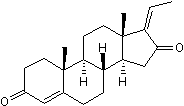 of an eight-bladed quinoprotein structure. As shown in Table 2, CcSDH shows some structural homology with known prokaryote PQQdependent sugar dehydrogenases, sGDH and soluble aldose sugar dehydrogenases from Pyrobaculum aerophilum and Escherichia coli, although the amino acid sequence homology is low. Among prokaryotic sugar dehydrogenases, CcSDH shows some structural similarity with human hedgehog-interacting protein. As seen in the alignment of the amino acid sequence of CcSDH with those of known six-bladed quinoproteins, a putative catalytic histidine is conserved in all proteins, whereas several amino acids contributing to PQQ binding in the prokaryote sugar dehydrogenase are missing in CcSDH and HHIP. However, as demonstrated above, the activity of CcSDH is PQQ-dependent. Thus, the amino acids involved in the adsorption of PQQ should be different in this enzyme family, indicating the diversity of PQQbinding motifs in eukaryotic enzymes. The PQQ-dependent sugar dehydrogenase that we discovered here has interesting enzymatic properties associated with characteristic cytochrome and cellulose-binding domains as well as the enzymes related to plant cell wall degradation, and shows high activity towards rare sugars. Therefore, the discovery of the PQQ-dependent domain in CcSDH could form the basis for a new AA family in the CAZy database. Detailed enzymatic studies will be necessary to evaluate why eukaryotes produce and secrete such enzymes, and how these enzymes acquire PQQ, because only a few prokaryotic organisms has been reported to be capable of synthesizing PQQ.
Author: KinaseInhibitorLibrary
By considering that a superior brake system decided by enabling fast approaches to turns
The apparent on/off functions of PTPN6 and CD3E may allow a T cell to initiate signaling events with maximal acceleration and then avoid deleterious overshoots through application of a molecular brake. Physiologically, we speculate that bang-bang control of TCR signal initiation may allow a T cell to launch rapid but controlled responses to infection. T cells scan antigen-presenting cells quickly and have been shown to decide if foreign antigen is present in under 1 min. Thus, the effect of a fairly short delay in phosphorylation of LAT or WAS, for example, could potentially have a major impact on the number of antigen-specific T cells responding to an infection. Bang-bang control is operative in gene circuits with negative autoregulation and in stem cell population dynamics and may represent a widely used design principle of cellular regulatory systems. Most cell surface glycans are highly sialylated and often involved in cell-cell and/or cell-extracellular matrix interaction. This involvement is mediated through I. their negative charge and II. by their interaction with specific Sia-binding proteins. Through this interaction  Sia play an important role in various biological processes such as growth, development, immunology, pathology and host-pathogen interactions. Aberrant expression of Ginsenoside-F4 sialylation has been observed as a characteristic feature for a variety of cancers, which enables cancer cells to escape from the immune surveillance and support these cells for increased migration and metastatic rates. Ligands for Sia-lectins, which are generated by sialyltransferases bind to Sia-lectins, such as selectins at the metastatic site and expand the cancer cell population. In addition, Sia also protect cancer cells from chemo- and radiation-therapy. Tumor cells of neural origin often express the neural cell adhesion molecule 1. This is known to be posttranslationally modified at the IgG domain V of N-glycans by polySia. PolySia have a unique structure of repeated units and can reach chain lengths of more than 50 sialic acid residues in mammals, which is primarily expressed on neural cell adhesion molecule 1. The biosynthesis of Sia starts in the cytosol. The physiological precursor of all Sia is N-acetylmannosamine. In a series of studies we provided evidence that the cellular Sia content can be metabolically engineered simply by feeding cells with synthetic Nacyl-modified D-mannosamines such as ManNProp or ManNPent. These synthetic Sia-precursors are taken up by the cells and are efficiently metabolized by the cellular sialylation machinery to the respective Sia. Neuroblastoma is one of the most frequent pediatric cancers, affecting children worldwide. The heterogeneity and complexity arose from various mutations. v-myc avian myelocytomatosis viral oncogene neuroblastoma derived homolog or anaplastic lymphoma receptor kinase, amplification and other genetic factors decide the aggressiveness and recurrence nature of neuroblastoma. Since neuroblastoma has restricted targets for the tumor specific immunotherapy, it is a challenge to develop a novel Dimesna strategy to treat aggressive neuroblastoma. Here we focused on targeting hyper-sialylation through MSE using neuroblastoma cells and modified Sia-precursors. We found that depending on the MSE strategy, sialylation and polysialylation of neuroblastoma cells are reduced and that as a consequence migration and invasion are decreased and the sensitivity towards anticancer drugs, such as 5-fluorouracil or cisplatin and radiation, is increased.
The HR2 is a conserved region predicted the TGFb induced EMT in ovarian cancers
Therefore, synergistically overexpression of KLF4 and inhibition of TGFb pathway will provide a novel approach in the developing new therapeutic drugs for the treatment of ovarian cancers. In summary, this is the first report showing that KLF4 functions as a tumor suppressor by inhibiting cell proliferation, migration and invasion in ovarian cancer cells through attenuating TGFbinduced EMT. The endoplasmic reticulum is a membranous organelle where Pancuronium dibromide proteins targeted for secretion or for the plasma membrane are folded and processed. Physiological conditions that impose large amounts of proteins in the ER, as for example, the production of insulin by pancreatic b-cells, may represent a challenge to the ER folding capacity. The Unfolded Protein Response is a homeostatic mechanism that attempts to balance the load of incoming proteins into the ER to its folding Folinic acid calcium salt pentahydrate capacity, to avoid the accumulation of toxic misfolded proteins, which otherwise would cause ER stress. In higher eukaryotes, the UPR has three signaling branches triggered by different ER transmembrane proteins: protein kinase -like ER kinase, activating transcription factor 6 and Inositol-requiring enzyme 1. Ire1 is conserved across all eukaryotes, presenting a luminal domain that detects the accumulation of misfolded proteins in the ER lumen, and a cytoplasmic domain with kinase and RNase activities that trigger downstream signaling. During ER stress, Ire1 is activated and catalyzes the unconventional splicing of an intron from X-box binding protein 1 mRNA or from its functional yeast homolog Hac1. This Ire1 mediated unconventional splicing event causes a frameshift during the translation of Xbp1 mRNA that introduces a new C-terminus with a potent trans-activation domain, generating an effective transcription factor. In addition to splicing of Xbp1 mRNA, Ire1 also cleaves a variety of mRNAs, mostly encoding proteins with signal peptide/ transmembrane domains that would represent an additional challenge to the ER folding machinery under ER stress. This mechanism was named RIDD and was also described in mammalian cells and in the fission yeast S. pombe. RIDD seems to be particularly important in cells undergoing very strong ER stress. The mechanism of targeting of a specific mRNA to RIDD seems to rely mostly on the existence of a signal peptide in its respective protein; the deletion of the signal peptide from known RIDD targets prevents their degradation and conversely, addition of a signal peptide to GFP is sufficient to promote the degradation of its mRNA by RIDD. One interesting exception is the mRNA encoding Smt3, a homologue of SUMO, which is cleaved by RIDD although it does not have a signal peptide in its sequence. Xbp1 also does not have a signal peptide in its  sequence and the mechanism of recruitment of the Xbp1 mRNA to the ER membrane is still unclear. Moreover, it seems that the mechanisms of recruitment to the ER membrane of Xbp1 mRNA in mammals and Hac1 mRNA in yeast are quite different. In yeast cells with no ER stress, the mRNA of unspliced Hac1 is found mostly in the cytoplasm, in association with stalled ribosomes. Upon ER stress, Hac1 mRNA is recruited to Ire1 clusters in the ER membrane, in a process that depends on a bipartite element present at the 39 untranslated region of the Hac1 mRNA. In contrast, ER membrane localization of mammalian Xbp1 is independent of the 39 untranslated region of Xbp1. Instead, the mRNA of Xbp1unspliced is translated under normal conditions and originates a polypeptide that associates with the membrane of the ER through two hydrophobic regions.
Hepcidin may directly suppress the generation of erythrocyte during inflammation
Here, our main focus was the effect of HCV/HIV-1 coinfection on iron status. We found that coinfected patients had significantly lower levels of serum iron and a lower Tfs than HCVmonoinfected patients; however, there were no differences between HCV/HIV-1-coinfected patients and healthy individuals. Even though the levels of serum hepcidin were lower in HCVmonoinfected and HCV/HIV-1-coinfected patients than in healthy individuals, the levels were higher in HCV/HIV-1coinfected patients than in HCV-monoinfected patients. HIV infects CD4+ helper T-cells, macrophages, and dendritic cells, all of which play vital roles in the immune response. Macrophages also play an important role in iron homeostasis by recycling around 30 mg of iron per day, which is 20�C30-fold greater than the amount absorbed from food. If macrophages are compromised by HIV, much of the iron may accumulate within the cells and is not released into the serum, resulting in reduced serum iron levels in infected patients. This is consistent with our data showing that the RBC was lower in coinfected patients than in monoinfected patients. The reason for this reduction in the RBC is complicated, and may involve the drugs taken by HIV-infected patients. For example, it is known that for infants, anemia prevalence and serum ferritin level decrease after HAART Cinoxacin without routine iron supplementation. All of HIV-infected patients in our study had received zidovudine-containing HAART treatment, either regularly or intermittently. The decreased iron load in HIVinfected patients may be caused by both of  HIV per se and HAART therapy. Despite the belief that HCV infection can be cured by consistent optimization of therapeutic regimens containing PEG-IFN/ Ribavirin, a certain proportion of chronically-infected patients do not respond to treatment. This may be due to different genotypes and single-nucleotide polymorphismsin the IL28B genes; however, dysregulated iron homeostasis may be another reason. Evidence suggests that excess iron exerts toxic and fibrogenic effects via pathways involving the production of oxyradicals and oxidative stress; indeed, several studies show that removing excess iron through therapeutic phlebotomy reduces the severity of hepatic inflammation associated with Riociguat BAY 63-2521 chronic HCV infection. Accordingly, determining the status of serum iron and other iron-associated parameters, will be helpful to understand the complexity of alternations in iron distribution in HCVinfected patients, particularly those coinfected with HIV-1. In the last decade it has become possible to annotate a greater number of lncRNA transcripts. Consequently the study of them has gained much significance as many of them have been linked with epigenetic, transcriptional and post-transcriptional regulation of gene expression. Despite having generally a lower level of concentration than protein coding transcripts, lncRNAs exhibit more tissue specific expressions. A vast set of lncRNA transcripts are differentially expressed during development where many of them play critical roles. LncRNAs are now known to have a major involvement in cancer. Though, till now, a majority of the lncRNAs have been linked with epigenetic modulation of gene expressions, they can also regulate gene expression by transcriptional or post transcriptional modes. LncRNAs can influence post-transcriptional regulation by interfering with the miRNA pathways, by acting as competing endogenous RNAs.
Transferrin mainly functions to regulate iron transport and to prevent excessive iron deposition
Many experimental and clinical studiessuggest that chronic iron deposition promotes the progression of liver damage and increases the risk of fibrosis, cirrhosis, and hepatocellular carcinomain chronic hepatitis Cpatients. Accumulating evidencesuggests that excess iron catalyzes the formation of highly reactive free radicals and hydroxyl radicals, which can damage lipids, Lithium citrate proteins and DNA. Liver satellite cells are susceptible to oxidative injury, and are easily transformed into collagen producing cells that contribute to the development of fibrosis. Furthermore, some studiessuggest that excess iron in the liver may have an adverse effect on a patient’s response to antiviral therapy. Many hypotheseshave been put forward to explain the accumulation of iron in the liver of patients with CHC, including hemochromatosis mutations, loss of iron from damaged hepatocytes, and HCV-induced disturbance of iron homeostasis. The mechanism underlying iron accumulation in HCV-infected livers is unclear; however, the recent discovery of hepcidin, a peptide hormone that regulates iron hemostasis, may provide some clues. Alterations in iron distribution are common in infectious diseases and many of these alterations may be attributable to actions of the iron-regulatory hormone hepcidin. Hepcidin degrades the sole cellular iron exporter ferroportin leading to reduced iron absorption in the intestine and iron retention in monocytes, macrophages and spleen.Several studiesreport increased or decreased iron levels in the serum of HIV patients. The liver is the main organ responsible for iron homeostasis, and the status of the liver is closely related to the distribution of iron within the body. Some iron-associated proteins, such as  hepcidin and transferrin, are primarily synthesized by hepatocytes. Oxysophocarpine Pathological iron overload is observed in.50% of chronically-infected HCV patients. This is important because increased hepatic iron storage is an independent risk factor for the development of HCC. Here, we found that serum iron and ferritin levels were higher in HCV-monoinfected patients than in healthy controls. Iron is stored in the liver in the form of ferritin. When the liver is damaged by viral infection, ferritin is released from the damaged hepatic cells and actively secreted by macrophages during inflammation as well. This leads to higher serum ferritin concentrations in HCV-infected patients. We also found that HCV-infected patients had lower serum concentrations of hepcidin and transferrin. Hepcidin is a 25-amino acid peptide hormone primarily synthesized by hepatocytes, which regulates iron homeostasis by controlling iron absorption in the gut, iron release from macrophages, and the recycling of iron through erythrophagocytosis. In agreement with the results presented herein, several studies report low hepcidin levels in HCV-infected patients. Hepcidin binds to ferroportin, a membrane iron exporter expressed at high levels on enterocytes and macrophages. This results in ferroportin internalization and degradation, and a subsequently reduction in iron levels in the plasma. Recent studies suggest that reduced serum hepcidin levels in patients with chronic hepatitis C are associated with higher Tfs and increased serum iron accumulation. HCV infection may directly modulate hepcidin expression, and that higher Tfs and increased iron accumulation result in resistance to hepcidin.